X-Ray
X-rays use radiation to quickly produce images of bones, joints, and soft tissue. Saint Luke's offers a broad array of high-quality services, whether for a suspected fracture, persistent cough, or swallowing issue. We offer convenient access and reliable results to help you get extraordinary care.
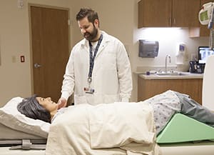
Having an X-ray
An X-ray uses a small amount of radiation to make images of your bones and organs. X-rays are most often used to look for bone or joint problems, or to check the heart and lungs (chest X-ray). They are also used to look for any hard deposits of minerals or salts in your body, such as kidney stones. You may also have an X-ray to check for problems in the bowel, such as a blockage or free air in the belly (abdomen). X-rays are also used to look for a foreign object in your body, such as metal. This can be from an injury or a swallowed object.
Tell the X-ray technologist
Tell them if you:
- Are or may be pregnant.
- Had an X-ray of this part of your body before.
- Have any metal objects on your body or clothes.
Before your test
Before the test starts, you may be asked:
- To remove your watch, jewelry, or clothes with metal closures from the part of your body being X-rayed. These items can block part of the image.
- To put on a hospital gown.
- About your overall health and any medicines you take.
During your test
Here is what to expect during the test:
- You may be asked to sit, stand, or lie on a table.
- A lead apron may be draped to protect the areas of your body not being X-rayed.
- With an X-ray of your chest or belly, you may have to take a deep breath and hold it for a few seconds.
- Each exam usually needs at least 2 X-ray views. You may need to move your body before each new X-ray view.
After your test
Your doctor will discuss the test results with you during a follow-up appointment or over the phone.
They may advise other imaging tests (such as an MRI or a CT scan) or more X-ray views if needed. This is done to confirm the diagnosis.