Duplex Ultrasound
Duplex ultrasound uses sound waves to get images of your blood vessels. It also helps determine the speed and the direction of blood flow through the vessels.
What Is Duplex Ultrasound?
Ultrasound is a test that uses sound waves to create detailed pictures of the inside of your body. Duplex ultrasound is a type of ultrasound that makes two kinds of images. First, it creates pictures of your blood vessels. Then, it makes graphs that show the speed and the direction of blood flow through the vessels. These images are viewed on a computer screen. No radiation or contrast fluid (dye) is used during the test. Ultrasound tests are safe to use during all stages of pregnancy.
What is duplex ultrasound used for?
Duplex ultrasound can help your healthcare provider find problems with blood vessels. These problems may include:
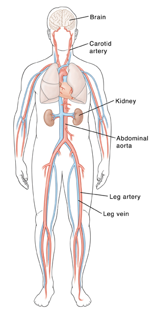
Carotid occlusive disease. These blocked or narrowed carotid arteries can lead to stroke.
Peripheral artery disease (PAD). These blocked or narrowed arteries in the arm or leg can cause pain.
Aneurysm. This is a ballooning out of a blood vessel wall.
Dissection. This is a tear in the layers of a blood vessel wall.
Deep vein thrombosis (DVT). This blood clot is in the deep veins of the arms or legs.
Varicose veins. These swollen, twisted veins can be seen under the skin’s surface.
Abnormal blood flow. This is an abnormal increase or decrease of blood flow to an area of your body.
Duplex ultrasound can also help your healthcare provider:
Decide whether further testing is needed
Determine the best treatment plan for you
Get more information about your blood vessels before surgery is done
What to Expect
Duplex ultrasound uses sound waves to get images of your blood vessels. It also helps determine the speed and the direction of blood flow through the vessels. Your doctor may want to do a duplex ultrasound to find out if you have any problems with the vessels that carry blood to and from the major organs in your body.

Before your test
Here's what you can expect:
- Follow instructions you are given to prepare for the test. If you are told to not eat or drink before the test, follow these directions carefully. If you don't, your test may need to be rescheduled.
- Tell your doctor what medicines you are taking. Ask if you should stop taking any of these medicines before the test. Also, bring a list of your medicines on the day of your test to show the technologist.
- Allow yourself enough time to check in.
What to tell the technologist
Tell the technologist if you’ve had:
- A previous procedure, such as a bypass, balloon angioplasty, or stent placement.
- Symptoms of stroke, including sudden short-term loss of strength, speech, or vision.
You may be asked other questions about your health. The information you give helps the technologist be sure the test is safe for you.
During your test
The duplex ultrasound is done in an ultrasound lab. The lab may be in your doctor’s office, a hospital, or an outpatient imaging center. The test is typically done by a vascular technologist. It can take from 15 minutes to 2 hours, depending on what’s being tested. During your test:
- You may be asked to change into a hospital gown or to remove clothing from the area being examined.
- You will likely lie on an exam table.
- A gel is applied to the area being tested. This helps sound waves move through your skin.
- The technologist slides the ultrasound probe (transducer) over the gel.
- You may hear a “whooshing” sound. This is the sound of your blood flowing. You may also see tracings of your blood flow on a screen.
- A blood pressure cuff may be put on your arm or leg and inflated during the test.
- You may be asked to stand up or even exercise during the test.
Be aware that
Be prepared for the following:
- The gel will feel wet. It won’t harm your skin or clothing, but it’s a good idea to wear washable clothes.
- The pressure from the probe may feel slightly uncomfortable. If you have any pain, tell the technologist.
After your test
Here is what to expect:
- Before leaving, you may need to wait briefly while your images are being reviewed.
- You can return to normal activities unless your doctor tells you not to.
- Your doctor will let you know when the test results are ready.
Locations
